Copper and Copper Oxides

Both the reddish-brown metal copper and also copper oxide are standard materials for electrical cables, for coins and an active ingredient in biocidal products (e.g. in wood preservatives and antifouling paints). Copper is also a common catalyst material. Due to its antimicrobial properties, copper oxide is found in consumer products such as pillowcases or socks. Copper is also an essential trace element and indispensable for important enzymes in the human body. Adults should consume approx. 1 – 1.5 mg daily.

Kupferkabel im Querschnitt. Bildquelle: Sergey-Ryzhov - stock.adobe.comHow can I come in contact with this material?

The risk of dermal uptake and skin sensitivity to copper (e.g. when handling of coins) is considered extremely low. However, copper oxide fumes can be breathed in and fume inhalation during the smelting of copper oxide powder can lead to metal fume fever, a disease with flu-like symptoms. Copper oxide can be found as a safe source of copper in over-the-counter vitamin supplements, but oral uptake of too high amounts of such supplements should be avoided.

Is there any risk from this material to humans and the environment?

Adverse effects in humans such as nausea and vomiting after the swallowing of copper oxide powder or lung damage after the breathing-in of copper oxide fumes have been reported. In most cases these effects are the result of exposure to very high doses of this metal and also to a higher bioavailability of copper with copper ions being released from copper oxide. However, copper is an essential trace element for the normal function of many tissues, including the nervous system, immune system, heart, skin as well as for the formation of capillaries. Copper is extremely well tolerated and processed by humans. This is not the case for all animals however and some wildlife species show greater sensitivity to copper toxicity.

Conclusion

Humans can have regular contact with copper and copper oxide through its many applications. It is important to keep in mind that copper oxide is a skin irritant and that its oral use (as an essential micro-element) should be limited. In addition, the handling of copper and copper oxide powder (also, as a nano-pesticide) should be done with great care.

Properties and Applications
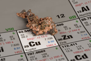
Kupfer auf Periodensystem der Elemente. Bildquelle: natros -stock.adobe.comCopper (chem. symbol Cu) is one of the world’s most important industrial metals. More than 20 million tons of Copper are consumed annually. This high demand, which is driven in particular by the industrial growth in Asia (62% of world copper consumption is in Asia), results from its excellent features.

This versatile metal is mainly used as an excellent heat and electricity conductor. Although for example silver and gold conduct similar well, but are much more expensive. Aluminum conducts electricity better than copper does, however, it is problematic to handle and in contrast to copper is not so inert. This meant that copper is the undisputed standard material for electrical wires. It occurs in almost all power cables, transformers, switches, processors and similar products.

Also Renewables cannot perform without the excellent conductivity of copper. With increasing electrification of our transportation (car, train, plane) also applications for copper are increasing. Recent high-speed trains such as the German ICE 3, for example, require 2 to 4 tons of copper – more than twice the amount of a normal electric powered train.

In construction, copper is also a popular raw material: Almost 60 percent of all German homes have water pipes made of copper. In addition, it is used as roofing material and is popular due to its resistance to extreme weather conditions. Copper forms in the presence of air a green patina on the surface, each one of us knows those roofs in particular of older buildings.

Besides its application as copper coinage, the biocidal agent is currently most widely used. As such substance is used in antifouling paints for ships. This results in the prevention of the growth of micro-organisms (fungi, algae etc.), whereby ships can theoretically save up to 40 percent fuel. For the preventive wood preservatives, industry takes advantage of this biocidal effect as well.

At the same time, copper is also an essential trace element. Adult humans require daily about 1-1,5 milligrams of the material. We take it on by copper-containing foods, such as chocolate, liver, cereals, vegetables and nuts.


Nano Copper

Streichen eines Gartenhauses aus Holz mit einem wasserfesten Holzschutzmittel zum Schutz vor Witterungseinflüssen. Bildquelle: David Pimborough - stock.adobe.comCopper Nanoparticles are mainly used as a biocide and for electrical applications. In the information and communication technology small, precise switching and conducting paths are increasingly needed. This is supported, for example, by solutions or pastes made ​​of copper nanoparticles, which can be selectively applied through novel printing process.

Antifouling coatings for ship hulls are also considered as application for copper nanoparticles. But currently no product can be identified, which made the statement ​​to use such particles as a biocide.

Unlike in the field of biocides for wood preservation: The wood is significantly improved by the use of copper nanoparticles in biocides, prolongs the life of wooden structures and thus significantly reduces the cost for the consumer. Aqueous formulations of copper nano – and microparticles in the order of 1 nm to 25 microns have been used for several years in the commercial wood preservation. The consumption of copper nanoparticles for this industry experts estimate that several thousand tons per year. Therby such wood preservatives have the potential to be one of the largest nano applications in Europe for hard woods such as Norway spruce (Picea abies) and silver fir (Abies alba).

Metallic nanocopper is self-igniting. The mixture of nanocopper with air (dust) is also ignitable without the influence of an ignition source. In contrast, nanoscale powder of copper oxide is not self- inflammable. Copper oxide dust just as little, so there is no possibility of a dust explosion in copper oxide ..


Occurrence and Production

In 2011, more than 16 million tons of copper from ore mines are mined. Already 5.3 million tons in Chile, currently the largest copper producer in the world. Copper is also good to recycle, so that even a large part of the global demand will be covered by so-called secondary copper.

Copper nanoparticles can be prepared by wet chemical methods or by flame synthesis. In the wet chemical variant copper salts, e.g. copper sulfate are reduced in the presence of a stabilizer (polymers, amine compounds, etc.) to nanoparticles. This one contains colloidal solutions of nanoparticles that can be processed directly. In flame synthesis under strongly reduced conditions cupreous compounds are burned, and so copper nanoparticles are obtained. There is the possibility of burning different metal-containing compounds, whereby hybrid metal particles (alloys) can be obtained.

Studies on copper and copper oxide particles show a toxic reaction due to the release of copper ions. High doses can cause various disorders in liver, kidney or neuronal functions due to the released copper ions.

Studies on Living Organisms – in vivo

Acute oral toxicity of copper particles has shown a significant correlation with its size distribution where micro-sized copper particles were classified as non-toxic (LD50 < 5000mg/kg). On the contrary, nano-sized copper particles had a LD50 of about 400mg/kg in mice and were classified as moderately toxic based on the Hodge and Sterner scale. After oral gavage of nano- and micro-particles with a concentration of 70mg/kg body weight in mice and an equivalent of soluble CuCl2 (147,6mg/kg) only the nano-copper generated an accumulation of excessive alkalescent substance and copper ions that even culminated in a metabolic alkalosis and overload of copper ions similarly to the results obtained with copper chloride ..

Another in vivo study conducted with rats analysing urine, serum, and extracts from liver and kidney tissues by metabolomic approach after up to 200mg Cu NP/kg body weight treatment for 5 days showed the induction of severe hepatotoxicity (toxin-induced liver disease) and nephrotoxicity (poisonous effect on the kidneys) similar to copper overload . The nephrotoxicity was mainly caused by an oxidative stress and at high doses (up to 600mg/kg body weight) cell death (apoptosis) via both extrinsic and intrinsic pathway was observed .

Treatment of mice via inhalation (4 hr/day 5d/week for 2 weeks with 3,5mg/m3) or instillation (24 hr post-exposure 3, 35, and 100µg/mouse) resulted in both cases in an induction of inflammatory responses and the recruitment of neutrophils into the lung. The simultaneous administration of Cu NP and bacteria impaired the pulmonary clearance against Klebsiella pneumonia .

After intranasal instillation of Cu-NP in mice not only the tissue and cells in direct contact are affected but also systemic effects have been observed. After middle dose of 40mg/kg body weight treatment significant changes in various neurotransmitter levels were measured in different brain regions, indicating a systemic effect in tissues were no particle or ion accumulation could be observed .

In general the described effects may be summarised in the following way: The particles determine the biodistribution and the release of copper ions, whereas the toxicity is comparable to cupper ions or other soluble nanomaterials.


Studies Outside of Organisms – in vitro

Studies of Cu and CuO particles have shown that these particles as others too tend to agglomerate quickly in cell culture medium. These agglomerates were taken up by different cell types in vesicular structures mostly and were not described to be free in the intracellular fluid (cytosol).

Cu and CuO nanoparticles as long as they are not coated are soluble in aqueous suspensions and release copper ions. Among the dose dependent cytotoxic effects, observed between 1-80µg/ml a gene expression study could demonstrate after treatment of A549 human lung carcinoma cells with 25µg/ml CuO nanoparticles (300nm agglomerates of 50 nm primary particle size) an up-regulation of metabolic processes and stress response genes as well as the down regulation of specific cellular processes and cell cycle regulating genes. In parallel the supernatant of CuO nanoparticles incubated cell culture medium was used for treatment experiments too and most of the effects observed with CuO nanoparticles could be seen as well, indicating that the CuO-NP toxicity is mainly due to the Cu ion release  .

In comparison with other nano-sized metal oxides such as TiO2, ZnO, Fe3O4, Fe2O3 or carbon-based materials e.g. carbon nanotubes, the non-coated nano-sized copper or copper oxide particles induced always the most severe cytotoxic effects at similar concentrations [3,4].

Several studies compared micro-sized with nano-sized CuO and showed independent of the cellular systems used e.g. A549, Hela or Chinese hamster oocytes, or CaCo-2 cells that the nano-sized material induces more severe effects in these cells due to their higher release of Cu ions.

Currently, there are no data regarding the environmental exposure to copper and copper oxide nanoparticles. In general, copper and copper compounds are naturally present in soils and waters.

Using computer models, a study has calculated that environmental concentrations of 0,06mg/l of copper nanoparticles could occur in Taiwanese rivers .

After inhalation or instillation of non-coated copper and copper oxide nanoparticles, no transfer into the bloodstream has been described to date. After oral administration, an increased level of copper ions was detected in the liver and kidney.

Uptake via the Lung – Inhalation

The determination of translocation across the air blood barrier is difficult for soluble particles since the dissolution of the particles may occur faster than the time point of analysis. However, as shown in the study of Zhang and coworkers, an increased level of copper ions was measured in liver, lung and olfactory bulb after inhalation of 40 mg/kg mice indicating that at least the released copper ions were able to cross biological barriers .


Uptake via the Gastro-Intestinal Tract

However, a confirmation of the presence of nanoparticles (NP) in the tissue is missing due to the solubility of copper (oxide) NPs. Copper (Cu) and copper oxide (CuO) particles induced hepatotoxic and nephrotoxic effects after application of high doses but the biological responses may be due to their Cu ion release .

There are numerous studies on the effect of copper (Cu) and copper oxide (CuO) nanoparticles on environmental organisms. Most take into account the effect of soluble copper ions and/or compare the effects of nanoscale Cu and CuOs with effects of coarser Cu and CuO particles. Some studies report a higher toxicity of nanoparticles compared to dissolved ionic copper but in many studies no differences in the effects could be found. Copper is one of the substances known to be ecotoxic, and copper compounds have been used for decades in paints for ship hulls to prevent the growth of algae, mussels and snails.

Copper and copper oxide nanoparticles were tested for their effects on different organisms living in different habitats. CuO nanoparticles are toxic to bacteria . However, copper ions dissolving from the particles are responsible for the effects; they interact with the "protective cover" of the bacteria and induce oxidative stress. In the presence of substances "scavenging" copper ions (chelators), no toxic effects were observed. Surprisingly, however, the toxic effects differed depending on the origin of ion: ions originating from particles act differently than ions from copper salts. Coarser particles released only very few ions and were not toxic.

The protozoan Tetrahymena responded to various copper compounds with a change in the composition of its fat components . At comparable toxicity nano-CuO released less ions compared to copper salts or coarse copper particles, hence an additional effect by the nano-form is assumed.


The effect of Cu nanoparticles on soil-dwelling worms was tested by mixing the particles into the soil. Depending on the worm species, contradictory results have been achieved. Cu nanoparticles had an inhibitory effect on the propagation of worms but administered in the same amounts as copper salt it was more toxic than the particles in one study and less toxic or of equal toxicity in another study. The effect of Cu nanoparticles and copper salts led to very different gene activation response patterns, leading to the assumption that the copper nanoparticles cause a specific effect, which is not comparable to copper ions .


Studies on zebrafish draw the same conclusion additionally showing a damage to the gills. Cu2O nanoparticles were less toxic for zebrafish than copper salts. Carp showed a growth delay after exposure to nano-CuO, a particle uptake into various organs was also detected .


CuO nanoparticles disrupt the development of Xenopus embryos. The main intake route was the ingestion of particles. Consequently, damage to the gastrointestinal tract was observed, which was due to both the particulate form and the ions. An electron microscopic study showed an uptake of CuO particles (nano and micro) in the intestines of water fleas but for both particle types no uptake from the gut into the body was observed. Water fleas showed a higher sensitivity to nanoparticles compared with coarser particles or salts .


A soil-dwelling water snail ingested more copper from nano copper oxide compared to coarser copper oxide and copper salts. Accordingly nano copper oxide had the greatest effects on growth, feed intake and reproduction of animals ..


Copper oxide particles had a toxic effect on green algae. In order to prevent the leaching of copper ions, CuO particles were coated with a polymer. These particles, however, showed a higher toxicity. The coating probably increases the particle uptake into the cells. Similar results were obtained on blue-green algae that took up more particles in the presence of organic materials. For various aquatic organisms (algae, crustaceans, rotifers) CuO particles were less toxic than copper salts, from the particles used in this study no leaching of ions was observed .


Different plant species (radish, ryegrass and duckweed) were inhibited in their growth in a consistent manner. In addition, DNA damage was observed in radish and ryegrasses. Although the absorption of copper in plants was higher when copper was present than ions, more damage in the genome occurred after exposure to nanoparticles. The duckweed took up more copper when it was present in particulate form. Corn plants showed nanoCuO induced growth retardation but not with Cu particles or coarse salt. A particle uptake by the roots and the distribution in the plant has been demonstrated [15]. Significant differences in the sensitivity of individual plant species were observed .


In summary, it can be stated that the known toxic effects of certain doses of copper occur likewise for the nano-form of copper. From the available studies no general statement can be made as to whether the effect is due solely to the ions or particulate form. The observed effects are due in part to the effect of the dissolving copper ions, and copper may be internalised in principle by many organisms. Between the two types of nanoparticles of CuO and Cu, there are no striking differences in the effects on organisms and there is no difference in the solubility.

Currently, there is no clear evidence that copper or copper oxide particles can pass biological barriers. They can be taken up by different cell types.

Behaviour at Tissue Barriers

Several ingestional or inhalation studies measured increased level of copper in different organs beyond the barrier tissue. However the analytical method applied is not always able to discriminate between nanoparticulate and ionic form. This is a general problem for (readily) soluble nanoparticles .


Behaviour of Uptake in somatic cells

Inside a cell the nanoparticles were accumulated in vesicular- or lysosomal-like structures.Uncoated particles will dissolve over the time in these organelles and induce either hot spots of high concentrations of Cu ions or may create hot spots of reactive oxygen species (ROS) formation .

Overall, there are very little data on the environmental behaviour. In a comparative study of several nanomaterials, nanoscale copper showed a relatively good mobility in artificial soil.

High salt concentrations resulted in a reduction of the mobility, whereas in the presence of humic acids, a slight increase was observed. Only few ions dissolute from the copper oxide nanoparticles used .

{4274171:ST2A8MSW},{4274171:JMCLSILS},{4274171:J8UBYZMX};{4274171:AYVWH7RL};{4274171:EACC9WLX};{4274171:CDV9X53A};{4274171:8VPXPUTM};{4274171:G6JLK6IV};{4274171:89CFWN3C},{4274171:MN4JBICZ},{4274171:WTIWXFM6},{4274171:FQ29FVPJ},{4274171:H8957M4K},{4274171:AKB67436};{4274171:GK8KLLFY};{4274171:8VPXPUTM},{4274171:G6JLK6IV};{4274171:FZ5SU7KQ},{4274171:EACC9WLX},{4274171:CDV9X53A};{4274171:7L32X59K},{4274171:Y6W4E8UD},{4274171:WMS47EX6};{4274171:YTX2ZJAQ};{4274171:YCDMNKYC},{4274171:4P8S7KCB},{4274171:HX9ASMTH};{4274171:YGUBNC9V},{4274171:2EEJKPEL},{4274171:5ZVVXSTT},{4274171:FI3V6QKM};{4274171:F7M9CTEH},{4274171:H85977JY},{4274171:IL48CSNW};{4274171:SQG8UDVQ};{4274171:IL48CSNW},{4274171:D4M8M33E},{4274171:2UT6MMD2},{4274171:EJHKXX83},{4274171:92E3Z8F8};{4274171:7MKK3V9S},{4274171:JDYU9GYH},{4274171:DMKMR22G};{4274171:EACC9WLX},{4274171:CDV9X53A},{4274171:G6JLK6IV};{4274171:89CFWN3C},{4274171:H8957M4K};{4274171:W9DJ97E3} default asc no 6664
1.
Lee, Y.; Choi, J.R.; Lee, K.J.; Stott, N.E.; Kim, D. Large-Scale Synthesis of Copper Nanoparticles by Chemically Controlled Reduction for Applications of Inkjet-Printed Electronics. Nanotechnology 2008, 19, 415604, https://doi.org/10.1088/0957-4484/19/41/415604.
1.
Evans, P.; Matsunaga, H.; Kiguchi, M. Large-Scale Application of Nanotechnology for Wood Protection. Nature nanotechnology 2008, 3, 577, https://doi.org/10.1038/nnano.2008.286.
1.
Magdassi, S.; Grouchko, M.; Kamyshny, A. Copper Nanoparticles for Printed Electronics: Routes Towards Achieving Oxidation Stability. Materials 2010, 3, 4626–4638, https://doi.org/Doi 10.3390/Ma3094626.
1.
Zhao, J.; Wang, Z.; Liu, X.; Xie, X.; Zhang, K.; Xing, B. Distribution of CuO Nanoparticles in Juvenile Carp (Cyprinus Carpio) and Their Potential Toxicity. Journal of hazardous materials 2011, 197, 304–310, https://doi.org/10.1016/j.jhazmat.2011.09.094.
1.
Wang, Z.; Xie, X.; Zhao, J.; Liu, X.; Feng, W.; White, J.C.; Xing, B. Xylem- and Phloem-Based Transport of CuO Nanoparticles in Maize (Zea Mays L.). Environmental science & technology 2012, 46, 4434–4441, https://doi.org/10.1021/es204212z.
1.
Zhang, L.; Bai, R.; Liu, Y.; Meng, L.; Li, B.; Wang, L.; Xu, L.; Le Guyader, L.; Chen, C. The Dose-Dependent Toxicological Effects and Potential Perturbation on the Neurotransmitter Secretion in Brain Following Intranasal Instillation of Copper Nanoparticles. Nanotoxicology 2012, 6, 562–575, https://doi.org/10.3109/17435390.2011.590906.
1.
Studer, A.M.; Limbach, L.K.; Van Duc, L.; Krumeich, F.; Athanassiou, E.K.; Gerber, L.C.; Moch, H.; Stark, W.J. Nanoparticle Cytotoxicity Depends on Intracellular Solubility: Comparison of Stabilized Copper Metal and Degradable Copper Oxide Nanoparticles. Toxicology letters 2010, 197, 169–174, https://doi.org/10.1016/j.toxlet.2010.05.012.
1.
Shi, J.; Abid, A.D.; Kennedy, I.M.; Hristova, K.R.; Silk, W.K. To Duckweeds (Landoltia Punctata), Nanoparticulate Copper Oxide Is More Inhibitory than the Soluble Copper in the Bulk Solution. Environmental pollution 2011, 159, 1277–1282, https://doi.org/10.1016/j.envpol.2011.01.028.
1.
Wang, Z.; Li, J.; Zhao, J.; Xing, B. Toxicity and Internalization of CuO Nanoparticles to Prokaryotic Alga Microcystis Aeruginosa as Affected by Dissolved Organic Matter. Environmental science & technology 2011, 45, 6032–6040, https://doi.org/10.1021/es2010573.
1.
Saison, C.; Perreault, F.; Daigle, J.C.; Fortin, C.; Claverie, J.; Morin, M.; Popovic, R. Effect of Core-Shell Copper Oxide Nanoparticles on Cell Culture Morphology and Photosynthesis (Photosystem II Energy Distribution) in the Green Alga, Chlamydomonas Reinhardtii. Aquatic toxicology 2010, 96, 109–114, https://doi.org/10.1016/j.aquatox.2009.10.002.
1.
Sarkar, A.; Das, J.; Manna, P.; Sil, P.C. Nano-Copper Induces Oxidative Stress and Apoptosis in Kidney via Both Extrinsic and Intrinsic Pathways. Toxicology 2011, 290, 208–217, https://doi.org/10.1016/j.tox.2011.09.086.
1.
Piret, J.P.; Vankoningsloo, S.; Mejia, J.; Noel, F.; Boilan, E.; Lambinon, F.; Zouboulis, C.C.; Masereel, B.; Lucas, S.; Saout, C.; et al. Differential Toxicity of Copper (II) Oxide Nanoparticles of Similar Hydrodynamic Diameter on Human Differentiated Intestinal Caco-2 Cell Monolayers Is Correlated in Part to Copper Release and Shape. Nanotoxicology 2012, 6, 789–803, https://doi.org/10.3109/17435390.2011.625127.
1.
Pang, C.; Selck, H.; Misra, S.K.; Berhanu, D.; Dybowska, A.; Valsami-Jones, E.; Forbes, V.E. Effects of Sediment-Associated Copper to the Deposit-Feeding Snail, Potamopyrgus Antipodarum: A Comparison of Cu Added in Aqueous Form or as Nano- and Micro-CuO Particles. Aquatic toxicology 2012, 106–107, 114–122, https://doi.org/10.1016/j.aquatox.2011.10.005.
1.
Perreault, F.; Oukarroum, A.; Melegari, S.P.; Matias, W.G.; Popovic, R. Polymer Coating of Copper Oxide Nanoparticles Increases Nanoparticles Uptake and Toxicity in the Green Alga Chlamydomonas Reinhardtii. Chemosphere 2012, 87, 1388–1394, https://doi.org/10.1016/j.chemosphere.2012.02.046.
1.
Mortimer, M.; Kasemets, K.; Vodovnik, M.; Marinsek-Logar, R.; Kahru, A. Exposure to CuO Nanoparticles Changes the Fatty Acid Composition of Protozoa Tetrahymena Thermophila. Environmental science & technology 2011, 45, 6617–6624, https://doi.org/10.1021/es201524q.
1.
Midander, K.; Cronholm, P.; Karlsson, H.L.; Elihn, K.; Moller, L.; Leygraf, C.; Wallinder, I.O. Surface Characteristics, Copper Release, and Toxicity of Nano- and Micrometer-Sized Copper and Copper(II) Oxide Particles: A Cross-Disciplinary Study. Small 2009, 5, 389–399, https://doi.org/10.1002/smll.200801220.
1.
Manusadzianas, L.; Caillet, C.; Fachetti, L.; Gylyte, B.; Grigutyte, R.; Jurkoniene, S.; Karitonas, R.; Sadauskas, K.; Thomas, F.; Vitkus, R.; et al. Toxicity of Copper Oxide Nanoparticle Suspensions to Aquatic Biota. Environmental toxicology and chemistry / SETAC 2012, 31, 108–114, https://doi.org/10.1002/etc.715.
1.
Meng, H.; Chen, Z.; Xing, G.; Yuan, H.; Chen, C.; Zhao, F.; Zhang, C.; Zhao, Y. Ultrahigh Reactivity Provokes Nanotoxicity: Explanation of Oral Toxicity of Nano-Copper Particles. Toxicology letters 2007, 175, 102–110, https://doi.org/10.1016/j.toxlet.2007.09.015.
1.
Lei, R.; Wu, C.; Yang, B.; Ma, H.; Shi, C.; Wang, Q.; Wang, Q.; Yuan, Y.; Liao, M. Integrated Metabolomic Analysis of the Nano-Sized Copper Particle-Induced Hepatotoxicity and Nephrotoxicity in Rats: A Rapid in Vivo Screening Method for Nanotoxicity. Toxicology and applied pharmacology 2008, 232, 292–301, https://doi.org/10.1016/j.taap.2008.06.026.
1.
Kim, J.S.; Adamcakova-Dodd, A.; O’Shaughnessy, P.T.; Grassian, V.H.; Thorne, P.S. Effects of Copper Nanoparticle Exposure on Host Defense in a Murine Pulmonary Infection Model. Particle and fibre toxicology 2011, 8, 29, https://doi.org/10.1186/1743-8977-8-29.
1.
Karlsson, H.L.; Gustafsson, J.; Cronholm, P.; Moller, L. Size-Dependent Toxicity of Metal Oxide Particles–a Comparison between Nano- and Micrometer Size. Toxicology letters 2009, 188, 112–118, https://doi.org/10.1016/j.toxlet.2009.03.014.
1.
Karlsson, H.L.; Cronholm, P.; Gustafsson, J.; Moller, L. Copper Oxide Nanoparticles Are Highly Toxic: A Comparison between Metal Oxide Nanoparticles and Carbon Nanotubes. Chemical research in toxicology 2008, 21, 1726–1732, https://doi.org/10.1021/tx800064j.
1.
Ivask, A.; Bondarenko, O.; Jepihhina, N.; Kahru, A. Profiling of the Reactive Oxygen Species-Related Ecotoxicity of CuO, ZnO, TiO2, Silver and Fullerene Nanoparticles Using a Set of Recombinant Luminescent Escherichia Coli Strains: Differentiating the Impact of Particles and Solubilised Metals. Anal Bioanal Chem 2010, 398, 701–716, https://doi.org/10.1007/s00216-010-3962-7.
1.
Heinlaan, M.; Kahru, A.; Kasemets, K.; Arbeille, B.; Prensier, G.; Dubourguier, H.C. Changes in the Daphnia Magna Midgut upon Ingestion of Copper Oxide Nanoparticles: A Transmission Electron Microscopy Study. Water research 2011, 45, 179–190, https://doi.org/10.1016/j.watres.2010.08.026.
1.
Hanagata, N.; Zhuang, F.; Connolly, S.; Li, J.; Ogawa, N.; Xu, M. Molecular Responses of Human Lung Epithelial Cells to the Toxicity of Copper Oxide Nanoparticles Inferred from Whole Genome Expression Analysis. ACS Nano 2011, 5, 9326–9338, https://doi.org/10.1021/nn202966t.
1.
Heckmann, L.H.; Hovgaard, M.B.; Sutherland, D.S.; Autrup, H.; Besenbacher, F.; Scott-Fordsmand, J.J. Limit-Test Toxicity Screening of Selected Inorganic Nanoparticles to the Earthworm Eisenia Fetida. Ecotoxicology 2011, 20, 226–233, https://doi.org/10.1007/s10646-010-0574-0.
1.
Gunawan, C.; Teoh, W.Y.; Marquis, C.P.; Amal, R. Cytotoxic Origin of Copper(II) Oxide Nanoparticles: Comparative Studies with Micron-Sized Particles, Leachate, and Metal Salts. ACS Nano 2011, 5, 7214–7225, https://doi.org/10.1021/nn2020248.
1.
Griffitt, R.J.; Weil, R.; Hyndman, K.A.; Denslow, N.D.; Powers, K.; Taylor, D.; Barber, D.S. Exposure to Copper Nanoparticles Causes Gill Injury and Acute Lethality in Zebrafish (Danio Rerio). Environmental science & technology 2007, 41, 8178–8186, https://doi.org/10.1021/es071235e.
1.
Griffitt, R.J.; Luo, J.; Gao, J.; Bonzongo, J.C.; Barber, D.S. Effects of Particle Composition and Species on Toxicity of Metallic Nanomaterials in Aquatic Organisms. Environmental toxicology and chemistry / SETAC 2008, 27, 1972–1978, https://doi.org/10.1897/08-002.1.
1.
Griffitt, R.J.; Hyndman, K.; Denslow, N.D.; Barber, D.S. Comparison of Molecular and Histological Changes in Zebrafish Gills Exposed to Metallic Nanoparticles. Toxicological sciences : an official journal of the Society of Toxicology 2009, 107, 404–415, https://doi.org/10.1093/toxsci/kfn256.
1.
Gomes, S.I.; Novais, S.C.; Scott-Fordsmand, J.J.; De Coen, W.; Soares, A.M.; Amorim, M.J. Effect of Cu-Nanoparticles versus Cu-Salt in Enchytraeus Albidus (Oligochaeta): Differential Gene Expression through Microarray Analysis. Comparative biochemistry and physiology. Toxicology & pharmacology : CBP 2012, 155, 219–227, https://doi.org/10.1016/j.cbpc.2011.08.008.
1.
Dimkpa, C.O.; Calder, A.; Britt, D.W.; McLean, J.E.; Anderson, A.J. Responses of a Soil Bacterium, Pseudomonas Chlororaphis O6 to Commercial Metal Oxide Nanoparticles Compared with Responses to Metal Ions. Environmental pollution 2011, 159, 1749–1756, https://doi.org/10.1016/j.envpol.2011.04.020.
1.
Chio, C.P.; Chen, W.Y.; Chou, W.C.; Hsieh, N.H.; Ling, M.P.; Liao, C.M. Assessing the Potential Risks to Zebrafish Posed by Environmentally Relevant Copper and Silver Nanoparticles. The Science of the total environment 2012, 420, 111–118, https://doi.org/10.1016/j.scitotenv.2012.01.023.
1.
Ben-Moshe, T.; Dror, I.; Berkowitz, B. Transport of Metal Oxide Nanoparticles in Saturated Porous Media. Chemosphere 2010, 81, 387–393, https://doi.org/10.1016/j.chemosphere.2010.07.007.
1.
Chen, D.; Zhang, D.; Yu, J.C.; Chan, K.M. Effects of Cu2O Nanoparticle and CuCl2 on Zebrafish Larvae and a Liver Cell-Line. Aquatic toxicology 2011, 105, 344–354, https://doi.org/10.1016/j.aquatox.2011.07.005.
1.
Bacchetta, R.; Santo, N.; Fascio, U.; Moschini, E.; Freddi, S.; Chirico, G.; Camatini, M.; Mantecca, P. Nano-Sized CuO, TiO(2) and ZnO Affect Xenopus Laevis Development. Nanotoxicology 2012, 6, 381–398, https://doi.org/10.3109/17435390.2011.579634.
1.
Atha, D.H.; Wang, H.; Petersen, E.J.; Cleveland, D.; Holbrook, R.D.; Jaruga, P.; Dizdaroglu, M.; Xing, B.; Nelson, B.C. Copper Oxide Nanoparticle Mediated DNA Damage in Terrestrial Plant Models. Environmental science & technology 2012, 46, 1819–1827, https://doi.org/10.1021/es202660k.
1.
Amorim, M.J.; Scott-Fordsmand, J.J. Toxicity of Copper Nanoparticles and CuCl2 Salt to Enchytraeus Albidus Worms: Survival, Reproduction and Avoidance Responses. Environmental pollution 2012, 164, 164–168, https://doi.org/10.1016/j.envpol.2012.01.015.
1.
Tang, M.; Zhang, T.; Xue, Y.; Wang, S.; Huang, M.; Yang, Y.; Lu, M.; Lei, H.; Kong, L.; Yuepu, P. Dose Dependent in Vivo Metabolic Characteristics of Titanium Dioxide Nanoparticles. Journal of nanoscience and nanotechnology 2010, 10, 8575–8583, https://doi.org/10.1166/jnn.2010.2482.
Skip to content